What is Abdominal Ultrasound Imaging?
Ultrasound imaging, also called ultrasound scanning or sonography, involves exposing part of the body to high-frequency sound waves to produce pictures of the inside of the body. Ultrasound exams do not use ionizing radiation (x-ray). Because ultrasound images are captured in real-time, they can show the structure and movement of the body's internal organs, as well as blood flowing through blood vessels.
Ultrasound imaging is usually a painless medical test that helps physicians diagnose and treat medical conditions.
An abdominal ultrasound produces a picture of the organs and other structures in the upper abdomen.
A Doppler ultrasound study may be part of an abdominal ultrasound examination.
Doppler ultrasound is a special ultrasound technique that evaluates blood as it flows through a blood vessel, including the body's major arteries and veins in the abdomen, arms, legs and neck

Jennifer A. McDowell, RT, RVT, RDMS

Abdominal Ultrasound
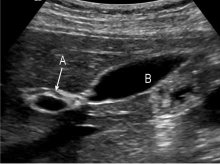
Gallbladder & Common Hepatic Duct

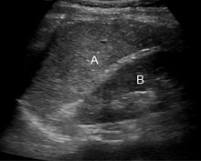
A-Spleen B-Kidney
Vascular Ultrasound
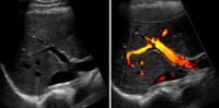
Main Portal Vein with and without blood flow

Normal aorta doppler

Ultrasound image of a kidney with normal blood flow

Yawning baby
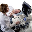
Ultrasound Machine
1 comment:
I love your blog and have some questions about the profession. How do I contact you with questions and are you available to answer them?
Post a Comment