Great News!
The abstract that I wrote with Dr. Romero about the use of 3-Dimensional Ultrasound on Neonatal Brains was accepted and I will be presenting it at the RSNA. The RSNA is a national conference in Radiology and is attended by 5-6,000 people in the imaging field. I am including the link to their website. Enjoy!!
http://rsna2007.rsna.org/rsna2007/V2007/index.cvn?id=10000&p_navID=262
Thursday, August 2, 2007
Monday, July 16, 2007
Ultrasound for infertility
Ultrasound technology provides a nonsurgical way of viewing a woman's pelvic organs during various infertility tests and procedures. It uses high-frequency sound waves that travel at different speeds through body organs and tissues. The waves are then reflected back to a detector where they are converted into pictures. The probe (transducer) that is used to assess and help treat infertility-related conditions is placed within the vagina (transvaginal).
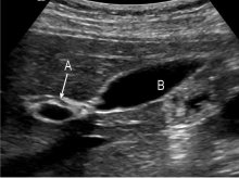
A hysterosonogram is done to evaluate the inside of the uterus (endometrial cavity) by filling the uterus with fluid during a transvaginal ultrasound. This procedure is also known as a sonohysterogram.
A hysterosonogram is done to evaluate the inside of the uterus (endometrial cavity) by filling the uterus with fluid during a transvaginal ultrasound. This procedure is also known as a sonohysterogram.
How Ultrasound Works
There are many reaons to get an ultrasound. Perhaps you are pregnant, and your obstetrician wants you to have an ultrasound to check on the developing baby or determine the due date. Maybe you are having problems with blood circulation in a limb or your heart, and your doctor has requested a Doppler ultrasound to look at the blood flow. Ultrasound has been a popular medical imaging technique for many years.
Monday, July 9, 2007
Prepare Yourself!
Proper patient preparation:
Abdominal Ultrasound-Nothing to eat or drink 8 hours prior to your exam.
Pelvic Ultrasound-Drink 16 ounces of clear liquid 1 hour prior to your exam.
Abdominal Ultrasound-Nothing to eat or drink 8 hours prior to your exam.
Pelvic Ultrasound-Drink 16 ounces of clear liquid 1 hour prior to your exam.
How should I prepare for the procedure?
You should wear comfortable, loose-fitting clothing for your ultrasound exam. You will need to remove all clothing and jewelry in the area to be examined.
You may be asked to wear a gown during the procedure.
Tell your doctor if you have had a barium enema or a series of upper GI (gastrointestinal) tests within the past two days. Barium that remains in the intestines can interfere with the ultrasound test.
Other preparations depend on the type of ultrasound you are having.
For a study of the liver, gallbladder, spleen, and pancreas, you may be asked to eat a fat-free meal on the evening before the test and then to avoid eating for eight to 12 hours before the test.
For ultrasound of the kidneys, you may be asked to drink four to six glasses of liquid about an hour before the test to fill your bladder. You may be asked to avoid eating for eight to 12 hours before the test to avoid gas buildup in the intestines.
For ultrasound of the aorta, you may need to avoid eating for eight to 12 hours before the test.
What is Abdominal Ultrasound Imaging?
Ultrasound imaging, also called ultrasound scanning or sonography, involves exposing part of the body to high-frequency sound waves to produce pictures of the inside of the body. Ultrasound exams do not use ionizing radiation (x-ray). Because ultrasound images are captured in real-time, they can show the structure and movement of the body's internal organs, as well as blood flowing through blood vessels.
Ultrasound imaging is usually a painless medical test that helps physicians diagnose and treat medical conditions.
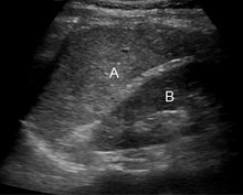
An abdominal ultrasound produces a picture of the organs and other structures in the upper abdomen.
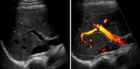
A Doppler ultrasound study may be part of an abdominal ultrasound examination.
Doppler ultrasound is a special ultrasound technique that evaluates blood as it flows through a blood vessel, including the body's major arteries and veins in the abdomen, arms, legs and neck
Ultrasound imaging, also called ultrasound scanning or sonography, involves exposing part of the body to high-frequency sound waves to produce pictures of the inside of the body. Ultrasound exams do not use ionizing radiation (x-ray). Because ultrasound images are captured in real-time, they can show the structure and movement of the body's internal organs, as well as blood flowing through blood vessels.
Ultrasound imaging is usually a painless medical test that helps physicians diagnose and treat medical conditions.
An abdominal ultrasound produces a picture of the organs and other structures in the upper abdomen.
A Doppler ultrasound study may be part of an abdominal ultrasound examination.
Doppler ultrasound is a special ultrasound technique that evaluates blood as it flows through a blood vessel, including the body's major arteries and veins in the abdomen, arms, legs and neck
Saturday, June 30, 2007
Volume ultrasound:
Useful tool for musculoskeletal, breast and pediatric imaging
Volume ultrasonography has become an established modality in some specialties during the last years. The 'state-of-the-art' high frequency volume ultrasound (HFVU), presently performed using electromechanical transducers, is based on the quick acquisition of a volume data set which can then reviewed and re-processed using various display formats.Despite the lack of fluid in the muscular system, the tool can be useful even for musculoskeletal imaging, affirms Dr Carlo Martinoli, Associate Professor of Radiology at the University of Genoa, who presented the findings of his initial experience with 3D-volume ultrasound during the European Congress of Radiology (ECR) on 10 March."HFVU offers better anatomical rendering of superficial structures", Dr. Martinoli said. "Multiplanar imagining not only improves the presentation of the US information but can also be used as a problem-solving technique for diagnostic benefit."
Subscribe to:
Posts (Atom)